
X-rays are one tool a doctor may use in diagnosing osteoarthritis. But X-rays may not always detect osteoarthritis, so a doctor will also consider other information.
Osteoarthritis occurs when cartilage wears down in a joint where bones meet, although
Read on to learn more about what osteoarthritis might look like on an X-ray, what to expect from an X-ray procedure, and what other tests doctors might use to diagnose hip osteoarthritis.
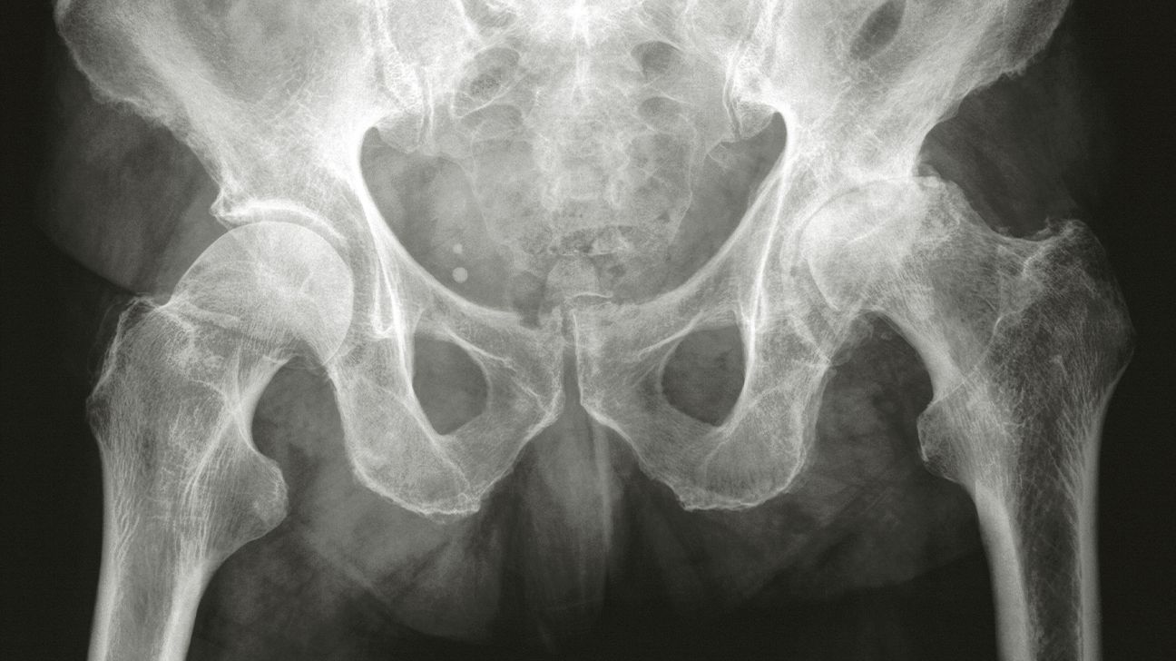
Your hip joint is a ball-and-socket joint. The ball-shaped, cartilage-covered femur head fits into a socket of the pelvic bone and allows your leg to move in many directions.
In a typical hip X-ray, you’ll see a space between the femur head and the pelvis where the cartilage cushions the femur in the joint.
If you have hip osteoarthritis, this joint space might look much narrower because the cartilage has worn away, allowing the femur head to move closer to the bone inside the pelvic socket.
This causes the bones to scrape against each other as you move your leg. When this happens, you might feel significant pain and stiffness when you try to walk, stand, or sit. You may also see cracks in the bone, pieces missing from the femur head, or white areas where the femur has hardened (subchondral sclerosis).
Pieces of damaged cartilage and bone from wear and tear to the joint may also be visible on a hip X-ray as white chunks around the joint. You may also notice bone spurs or cysts that have grown on your bone surfaces due to disease in the joint.
An X-ray may also show chondrocalcinosis, the buildup of calcium crystals in the joints. This
An X-ray can show wear and damage in the hip that results from osteoarthritis.
A hip X-ray is often able to show:
- how much the cartilage has worn down
- how narrow the joint space is
- how damaged the femur head is
Still, an X-ray may not always show cartilage damage from osteoarthritis.
Radiology labs or clinics that specialize in imaging tests typically perform hip X-rays.
You don’t need to do much to prepare for a hip X-ray. Here are some tips to help make the X-ray process more comfortable:
- Wear loose clothing that’s comfortable and easy to take on and off if you have to change into a gown.
- Take off any jewelry or metal accessories that may stop X-rays from producing clear images.
- Let the technician know if you have any metal implants in your body that may interfere with X-ray results.
Some X-rays will require standing next to a plate-shaped tool that can produce X-ray images. Other X-rays may require lying down so the technician can move a specialized camera over your hip to take X-ray images.
Stand or lie very still as the X-ray image is taken. This helps make sure that the image is clear. Once the technician gets the needed images, you can change into your regular clothes and leave the facility shortly afterward.
You may be able to review X-rays with a radiologist or doctor right away. But you may need to schedule a follow-up appointment to discuss the results and what treatments you might need.
X-ray images can’t always confirm a diagnosis of hip osteoarthritis, especially early on, according to 2022 research.
On an X-ray, doctors can’t always detect early structural changes, such as joint space narrowing and damage to the femur head. This is often when you’ll first start experiencing noticeable pain and changes in your walk. Many people who experience this pain don’t have any signs that show up on an X-ray.
Doctors need to use X-rays and other diagnostic tests to see how far along your osteoarthritis is and how much damage there is to the joint. This can help them choose an appropriate treatment option, such as hip arthroplasty.
Some other tests and procedures a doctor may use to detect hip osteoarthritis include:
- physical exam
- discussion about your symptoms, such as your pain levels and your movement
- a review of your
risk factors , such as age, weight, and family history - a comparison of your
leg lengths - a close inspection of how you walk
- additional imaging, such as magnetic resonance imaging (MRI) or computed tomography (CT), to look at the nerves and blood vessels around the joint
- blood tests to look for signs of rheumatoid arthritis (RA)
Doctors usually perform hip X-rays and other diagnostic tests to look for signs of wear to the hip.
Osteoarthritis is relatively easy to treat if caught early. If you’re experiencing a lot of pain and stiffness when you walk, contact a doctor to get X-ray imaging and other tests and learn what next steps you should take to treat the cause.