
Imaging like MRI and CT scans can help doctors detect signs of Crohn’s disease, like bowel wall thickening or ulcers. Ultrasounds and X-rays may also be of use.
Crohn’s disease is a chronic bowel disease that causes inflammation along your digestive tract. Experts collectively classify it as an inflammatory bowel disease (IBD) with ulcerative colitis.
The
- a physical exam
- lab tests
- imaging
Imaging tests play an essential role in:
- ruling out other conditions
- assessing the extent of your disease
- monitoring you for complications
This article explains why and when doctors use these imaging techniques and what signs they look for that may indicate Crohn’s disease.
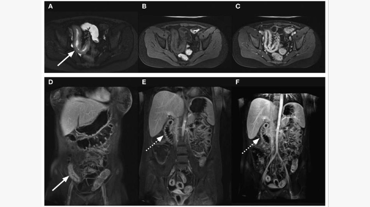
An MRI is the gold standard imaging technique for viewing patches of inflammation that indicate Crohn’s disease. They can also help a doctor notice complications and the extent of your disease activity.
Features of Crohn’s disease that may show up on an MRI include:
- thickened bowel walls
- strictures
- ulcerations
- creeping fat
- skip lesions
Crohn’s disease often requires repeated scans of your intestines to track how your disease changes over time. Unlike CT scans and X-rays, an MRI doesn’t expose your body to radiation.
The MRI techniques doctors use to visualize Crohn’s include:
- MR enteroclysis: During this procedure, you receive a tube through your nose that leads to your small intestines. A healthcare professional then injects a contrast solution through this tube.
- MR enterography: During this procedure, you drink a fluid that fills your bowel. This can help your doctor monitor disease activity and the effectiveness of treatments such as immune-modulating agents.
In a
- diagnosing small bowel disease extent (80% versus 70%)
- detecting disease presence (97% versus 92%)
- correctly ruling out small bowel disease extent (95% versus 81%)
- correctly ruling out disease presence (96% versus 84%)
Bhargava R, Hahn G, Hirsch W, Kim MJ, Mentzel HJ, Olsen OE, Stokland E, Triulzi F, Vazquez E. Libertas Academica Ltd. CC-BY-NC 3.0
A CT scan uses a series of X-rays to create a detailed image of your abdomen. Doctors use them to assess Crohn’s disease:
- activity
- extent
- complications
Features of Crohn’s disease that show up on a CT scan include:
- thickened bowel walls
- creeping fat
- “
comb sign ” showing the swelling of blood vessels in one of the bowel layers
A doctor may recommend a conventional CT scan with or without a contrast dye injection if you have a
CT enterography is a type of CT scan that can better visualize your small intestines than a conventional scan. This procedure involves drinking a special contrast liquid that shows up in the scanner. CT scans can accurately identify or rule out Crohn’s disease in more than
Copyright © 2013 Korean Society of Gastrointestinal Endoscopy CC BY-NC 3.0
Ultrasound is a radiation-free type of imaging that uses sound waves to produce an image of your gastrointestinal tract. It can also help identify health complications of other organs, such as your:
- liver
- bladder
- gallbladder
Abdominal ultrasounds are quick and widely available. They
- whether your disease is in remission
- potential complications
- disease severity
Ultrasounds can correctly identify Crohn’s inflammation around
Copyright © 2016 Rune Wilkens et al. CC BY 4.0
X-rays have a
- ulcers
- blockages
- fistulae
A barium enema involves putting a contrast dye into your anus with a tube so your doctors can better visualize your bowel on an X-ray.
Doctors don’t frequently use barium enemas. They more often perform CT scans or colonoscopies to identify changes to your colon.
Mediscan / Alamy Stock Photo
Imaging can help your doctor understand the extent of your Crohn’s disease and screen you for complications. Many experts consider an MRI the most accurate imaging tool for assessing Crohn’s, but CT scans and ultrasounds can make quicker and cheaper alternatives.
| Imaging | MRI | CT | Ultrasound | X-ray |
|---|---|---|---|---|
| Usage | assessing disease activity, extent, and complications | assessing disease activity, extent, and complications | assessing disease activity, severity, and complications | assessing complications |
| Price | expensive | moderately expensive | cheap | cheap |
| Speed | slow | fast | fast | fast |
| Accuracy | higher accuracy for detecting disease activity than ultrasound | comparable to MRI | less accurate than MRI but still highly accurate | limited use |
| Radiation exposure | no | yes | no | yes |